
Microscopy lets us take a much, much closer look at the details we might never see with the naked eye, and some multi-talented scientists know how to work a camera and editing suite. Throw those elements together and you’ve got one heck of a photo competition.
From a tiny turtle embryo that looks like a Tamagotchi to the most stunning image of an ovary we’ve ever seen, 20 images have been selected as the winners of the 45th Nikon Small World competition, which recognises excellence in photography taken under the microscope.
It’s a pretty niche type of photography (and you need the right equipment and access to the subjects), but the competition received over 2,000 entries from scientists in around 100 countries.
The competition was first launched in 1974, and judges entrants on originality, visual impact, informational content, and of course, technical proficiency. Here are the top 20 place-winners, starting with the winners, then in order of our favourites.
First place
The top spot was landed by microscopy technician Teresa Zgoda and university graduate Teresa Kugler with their incredible image of a turtle embryo, captured using fluorescence and stereo microscopy.

Look at this incredible turtle embryo.
Image: Teresa Zgoda & Teresa Kugler, Campbell Hall, Fluorescent turtle embryo, Stereomicroscopy, Fluorescence. 5x (Objective Lens Magnification)
Second place
In second place, Dr. Igor Siwanowicz, who used confocal microscopy to shoot a composite image of three single-cell freshwater protozoans, sometimes called “trumpet animalcules.”
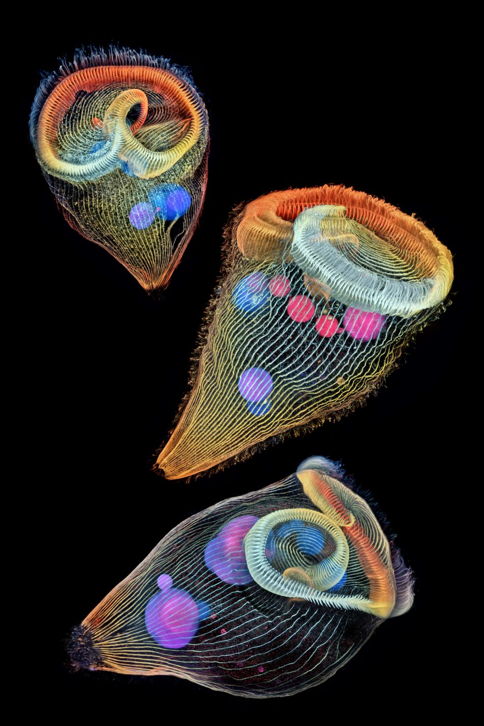
Teeny tiny single-cell freshwater protozoans.
Image: Dr. Igor Siwanowicz, Howard Hughes Medical Institute, Janelia Research Campus Ashburn, Depth-color coded projections of three stentors (single-cell freshwater protozoans), Confocal, 40x (Objective Lens Magnification)
Third place
In third place, Daniel Smith Paredes, with a pretty damn stunning image of a developing American alligator embryo, snapped at around 20 days of development using immunofluorescence.
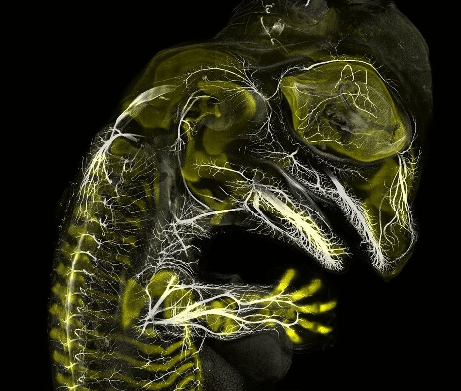
It’s a baby alligator!
Image: Daniel Smith Paredes & Dr. Bhart-Anjan S. Bhullar, Yale University, Department of Geology and Geophysics, Alligator embryo, developing nerves and skeleton, Immunofluorescence, 10x (Objective Lens Magnification)
And the other incredible place-winners:
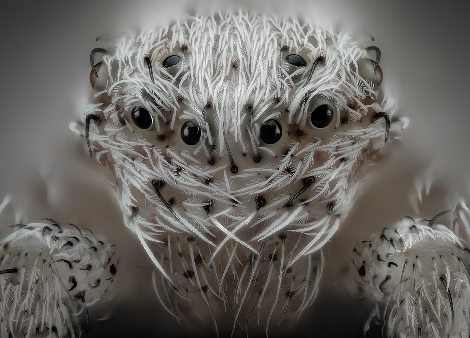
Just LOOK at this spider.
Image: JAVIER RUPÉREZ, ALMÁCHAR, MÁLAGA, SPAIN, SMALL WHITE HAIR SPIDER, REFLECTED LIGHT, IMAGE STACKING, 20X (OBJECTIVE LENS MAGNIFICATION)

Have you ever seen a tulip from this angle?
Image: Andrei Savitsky, Cherkassy, Ukraine, Tulip bud cross section, Reflected Light, 1x (Objective Lens Magnification)
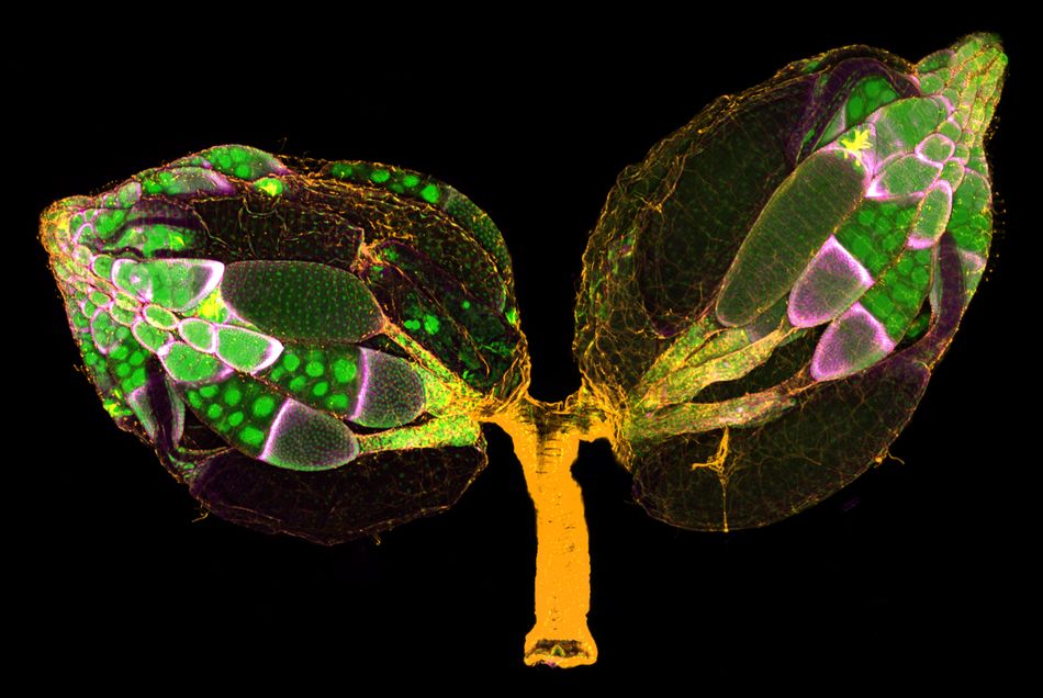
THAT is a pair of ovaries. RESPECT.
Image: Dr. Yujun Chen & Dr. Jocelyn McDonald, Kansas State University, Department of Biology, Manhattan, Kansas, USA, A pair of ovaries from an adult Drosophila female stained for F-actin (yellow) and nuclei (green); follicle cells are marked by GFP (magenta), Confocal, 10x (Objective Lens Magnification)
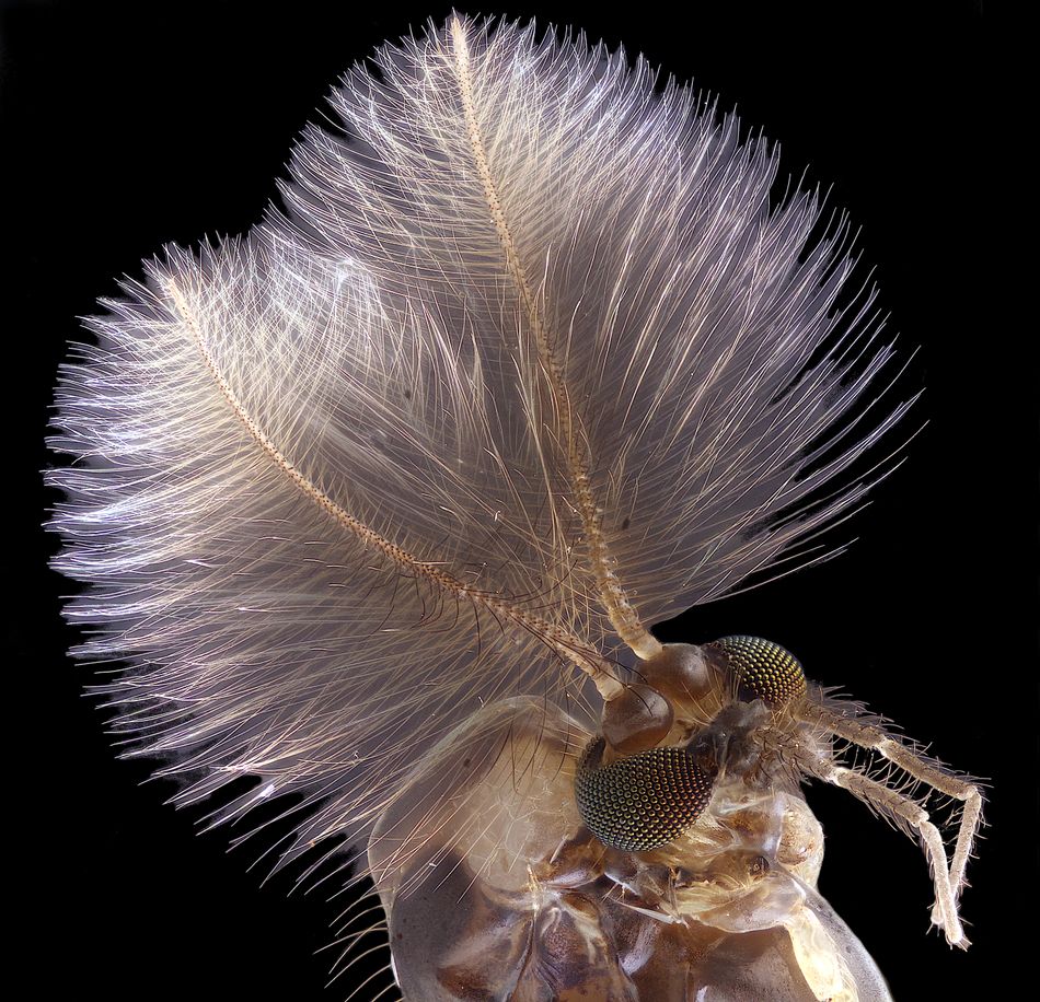
No one wants to be this close to a mosquito, even if it has awesome hair.
Image: Jan Rosenboom, Universität Rostock, Rostock, Mecklenburg Vorpommern, Germany, Male mosquito, Focus Stacking, 6.3x (Objective Lens Magnification)
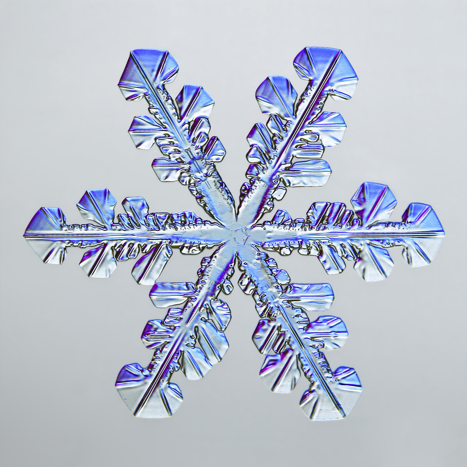
No two are the same.
Image: Caleb Foster, Caleb Foster Photography, Jericho, Vermont, USA, Snowflake, Transmitted Light, 4x (Objective Lens Magnification)

Looks like Elsa’s work.
Image: Garzon Christian, Quintin, Cotes-d’Armor, France, Frozen water droplet, Incident Light, 8x (Objective Lens Magnification)
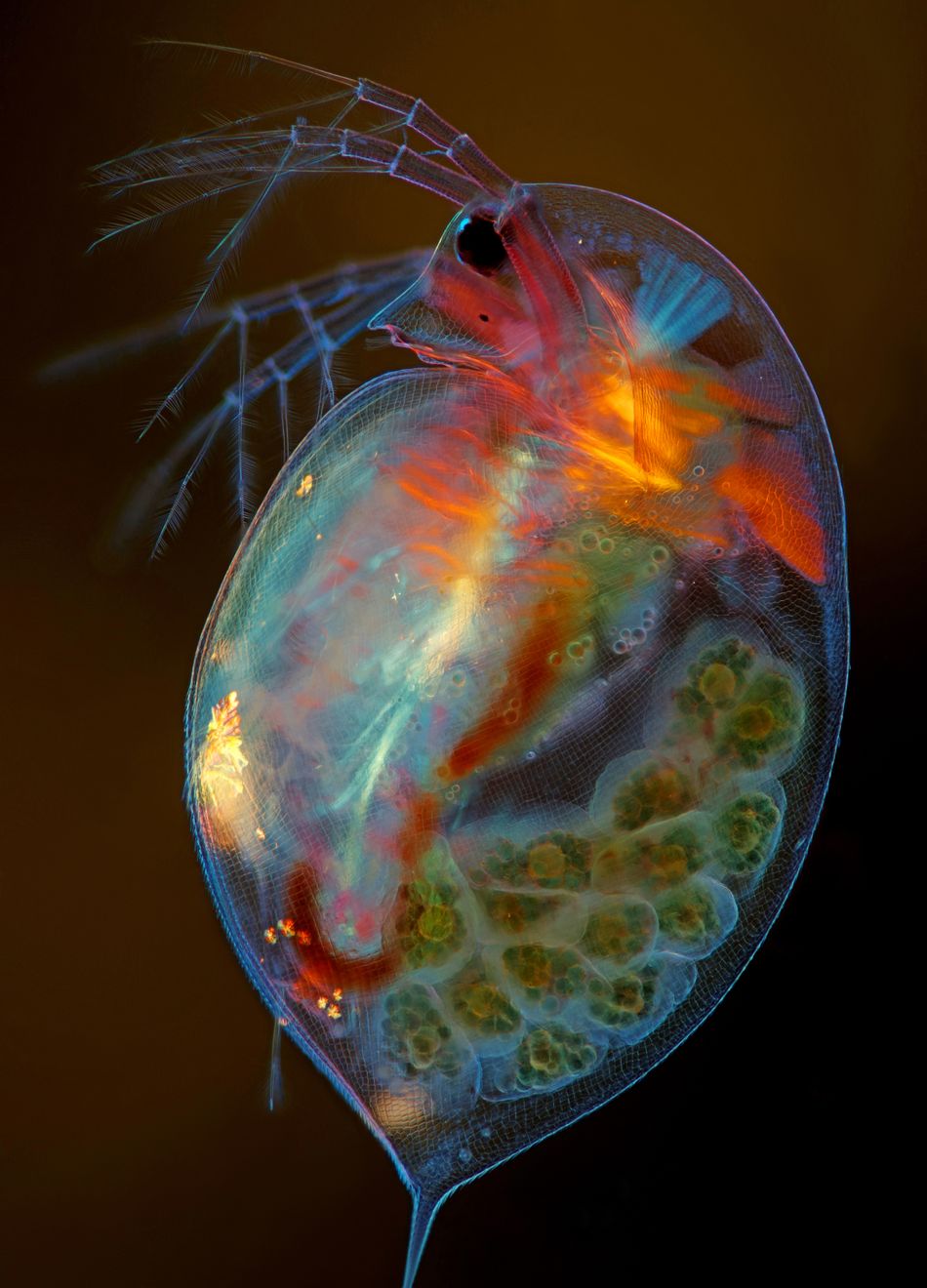
This small planktonic crustacean is pregnant.
Image: Marek MiśA160, Marek Miś Photography, Suwalki, Podlaskie, Poland, Pregnant Daphnia magna (small planktonic crustacean), Modified Darkfield, Polarized, Light, Image Stacking, 4x (Objective Lens Magnification)

A disco-hued octopus embryo.
Image: Martyna Lukoseviciute & Dr. Carrie AlbertinA202, University of Oxford, Weatherall Institute of Molecular Medicine, Oxford, Oxfordshire, United Kingdom, Octopus bimaculoides embryo, Confocal, Image Stitching, 5x (Objective Lens Magnification)
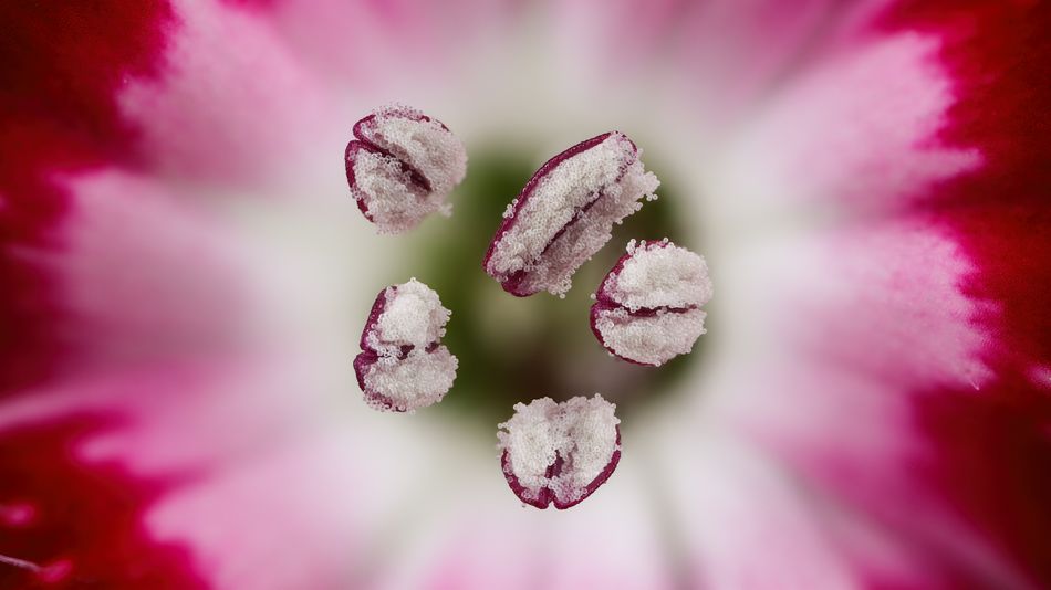
Preeeetty.
Image: Dr. Guillermo López López, Alicante, Spain, Chinese red carnation stamen, Focus Stacking, 3x (Objective Lens Magnification)

Make sure you get up close to your daily vitamin C.
Image: arl DeckartA181, Eckental, Bavaria, Germany, Vitamin C, Brightfield, Polarized Light, 4x (Objective Lens Magnification)

Teeny baby mosquito.
Image: Anne Algar, Hounslow, Middlesex, United Kingdom, Mosquito larva, Darkfield, Polarizing Light, Image Stacking, 4x (Objective Lens Magnification)A133
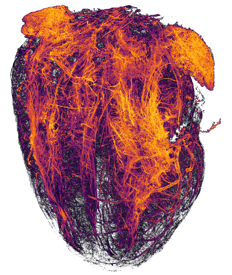
Be still, my little beating mouse heart.
Image: Simon Merz, Lea Bornemann & Sebastian KorsteA214, University Hospital Essen, Institute for Experimental Immunology & Imaging, Essen, Nordrhein-Westfalen, Germany, Blood vessels of a murine (mouse) heart following, myocardial infarction (heart attack), Tissue Clearing, Light Sheet, Fluorescence Microscopy, 2x (Objective Lens Magnification)
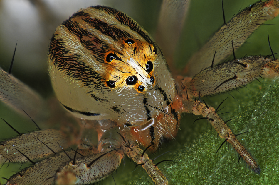
Oh heyyyyy another spider.
Image: Antoine Franck, CIRAD – Agricultural Research for Development, Saint Pierre, Réunion, Female Oxyopes dumonti (lynx) spider, Focus Stacking, 1x (Objective Lens Magnification)
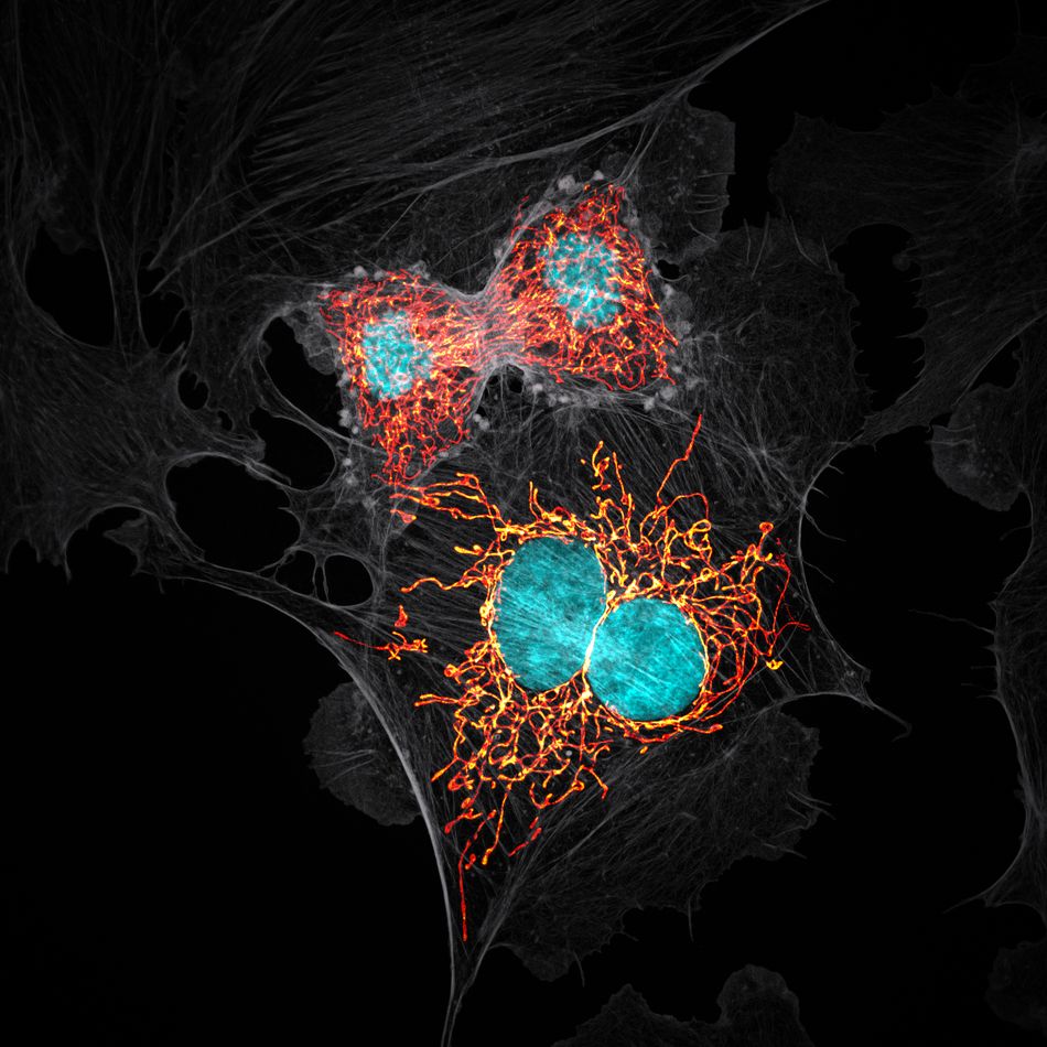
Get all up in this moment of mitosis.
Image: Jason M. Kirk, Baylor College of Medicine, Optical Imaging & Vital Microscopy Core, Houston, Texas, USA, BPAE cells in telophase stage of mitosis, Confocal with Enhanced Resolution, 63x (Objective Lens Magnification)

This close-up of cuprite looks like Minecraft.
Image: Dr. Emilio Carabajal MárquezA139, Madrid, Spain, Cuprite (mineral composed of copper oxide), Focus Stacking, 20x (Objective Lens Magnification)
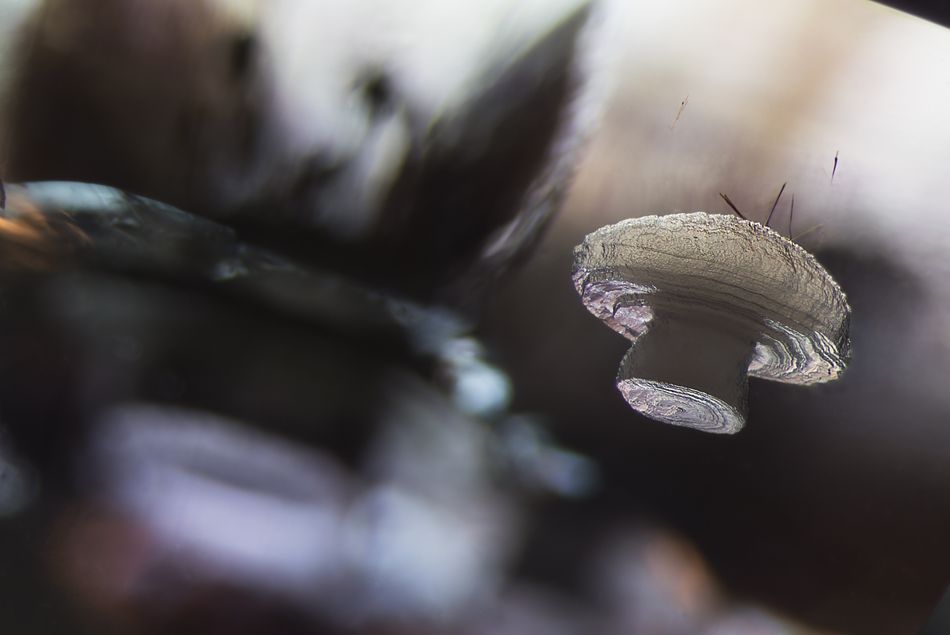
That’s not a mushroom, it’s a cristobalite crystal.
Image: E. Billie HughesA191, Lotus Gemology, Bangkok, Thailand, Cristobalite crystal suspended in its quartz mineral host, Darkfield, 40x (Objective Lens Magnification)
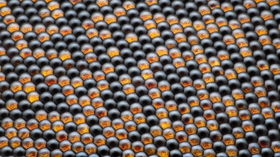
That’s the eye of a fly.
Image: Dr. Razvan Cornel ConstantinA171, Bucharest, Romania, Housefly compound eye pattern, Focus Stacking, Reflected Light, 50x (Objective Lens Magnification)
Want more brilliant photography involving living things? Want it to be… funny? Right this way.
read more at https://mashable.com/?utm_campaign=Mash-Prod-RSS-Feedburner-All-Partial&tm_cid=Mash-Prod-RSS-Feedburner-All-Partial by Shannon Connellan
Digital marketing


